Name: Textbook of Orthopaedics
Edition: 5th
Author: John Ebnezar; Rakesh John
Subject: Orthopaedics
Language: English
Publisher: Jaypee Brothers Medical Publishers

Brief Introduction
Highlights of Textbook of Orthopaedics
• The book has been thoroughly overhauled
• Each and every chapter has been thoroughly updated
• I have inserted about 1,350 mind-boggling color diagrams in this edition
• I have inserted a number of color clinical photographs wherever required
• New X-rays have been added
• Breaking away from the conventional practice, I have introduced lots of innovative and thought-provoking diagrams, which is an idea of my own. Hope students will like it
• Even the appendices have been thoroughly revised
• Due to the sheer number of diagrams, the book has slightly overgrown its size. However, it is an apparent bulk and not the real bulk as diagrams add to the quality of the book.
Contents
Section 1: Traumatology
- Trauma—A Modern International Epidemic
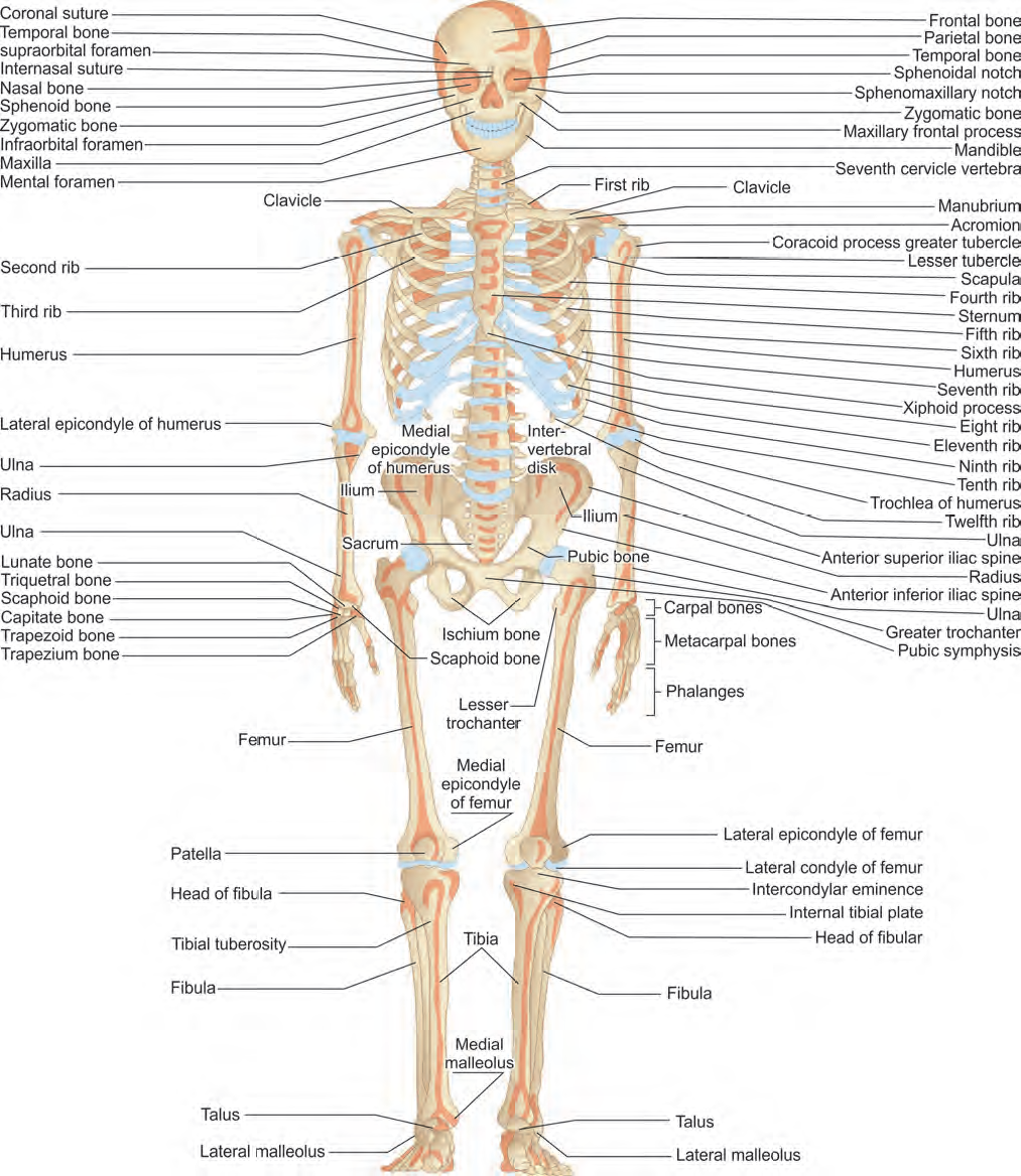
- Know Your Skeletal System
- General Principles of Fractures and Dislocations
- Complications of Fractures
- Emergency Care of the Injured
- Fracture Treatment Methods: Then, Now and Future
- Recent Advances in Fracture Treatment
- Fracture Healing Methods
- Soft Tissue Injuries
- Fractures in Special Situations
- Injuries Around the Clavicle
- Injuries of the Shoulder Joint
- Injuries Around the Elbow
- Injuries of the Forearm
- Injuries to the Wrist
- Hand Injuries
- Dislocations of the Hip Joint
- Fracture Femur
- Injuries of the Knee
- Fracture of Tibia and Fibula
- Injuries of the Ankle
- Injuries of the Foot
- Pelvic Injuries, Rib and Coccyx Injuries
- Injuries of the Spine
- Peripheral Nerve Injuries
- Approach to Orthopedic Disorders
- Deformities and their Management
- Treatment of Orthopedic Disorders
- Regional Conditions of the Neck
- Regional Conditions of the Upper Limb
- Regional Conditions of the Spine
- Regional Conditions of the Lower Limb
- Disorders of the Hand
- Low Backache and Repetitive Stress Injury
- Congenital Disorders
- Developmental Disorders
- Metabolic Disorders
- Osteomyelitis
- Skeletal Tuberculosis
- Disorders of Joints (Arthritis)
- Rheumatic Diseases
- Neuromuscular Disorders
- Bone Neoplasias
- Distal Forearm Fractures
- Fracture Neck of Femur
- Osteoporosis
- Osteoarthritis
- Cervical Disk Syndromes
- Degenerative Lumbar Disk Disease and Canal Stenosis
- Common Surgeries of the Humerus
- Common Forearm Surgeries
- Common Hip Surgeries
- Common Surgery of the Femur
- Common Surgery of the Patella
……..
Clinical Examination Methods in Orthopedics
- Examination of a Bony Swelling
- Examination of Shoulder Joint
- Examination of Elbow Joint
- Examination of Wrist
- Examination of Hip Joint
- Examination of Knee Joint
- Examination of Sacroiliac Joint
- Examination of Spine
Excerpts
…
Bone makes a valiant attempt to get back to its original shape and form after having suffered humiliating fractures due to a myriad of incriminating forces. Bone is unique in healing itself completely with a tissue that is indistinguishable from the original tissue hence there is no scar left. The term bone regeneration and not fracture healing is more appropriate. Bone is repaired by callus, which is a new tissue that may develop externally or internally. An external callus envelops around the outer aspect of the opposing ends of bone fragments. An internal callus forms between the bone ends. During the first two days at the fracture site and away from the fracture site, in the deep layer of the periosteum the osteogenic cells proliferate and lift the fibrous layer of the periosteum away from the bone. Marrow cells also proliferate but to a lesser degree. These osteogenic cells differentiate into osteoblasts, which form the bone trabeculae resembling the embryonic tissue. The osteogenic cells lying away from the fracture site due to inadequate vascularity differentiate into chondroblasts and chondrocytes, which form the cartilage. The cartilage is finally converted into bone by endochondral ossification.
Page 26
…
Screenshots


Download Links
The download links are hidden, you should sign-in and pay the fee(diamonds) or points to get the links.